What is diagnostic ultrasound (US)?
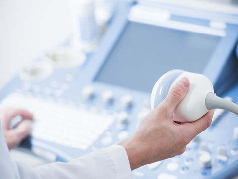
An ultrasound transducer sends a beam of ultrasonic waves to the body. At the boundary of two substances with different density, these waves are reflected back into the transducer where they are detected. An image on the screen of the scanner is then created by computer processing.
US allows the examination of superficial tissues with very high spatial resolution. It does not pass through the bone, so other changes in bone, bone marrow, and structures that lie within the joints, except on its surface, cannot be imaged.
Unlike other radiological investigations, US allows for a dynamic examination. Namely, certain pathological changes are not apparent or become more pronounced only when the muscle is contracted or at certain movements. The possibility to compare symmetrical structures on the healthy and injured or diseased sides is an additional advantage. Doppler ultrasound allows a very sensitive presentation of tissue vascularization and flow in large vessels. US is of great value in the guidance of puncture needles for diagnostic and therapeutic purposes. Moreover, it has no known adverse health effects.
Does the patient have to be prepared for the examination?
To ensure optimal transparency, do not eat anything (for approximately 6 hours) before an ultrasound investigation of the abdominal organs. For the urinary tract examination, drink 0.5 to 1 liter of water one hour before the procedure.
No special preparation is required for the ultrasound investigation of joints and soft tissues, as well as for the ultrasound of veins.
The clothing in the examined part must be taken off for the process.
How is the procedure performed?
The patient lies or sits on the examination table, depending on the type of the investigation. The radiologist guides the probe over the part of the body to be examined and observes the changes on the screen.
The process takes about 15 minutes.
This investigation is painless.
US examinations are performed exclusively on a self-pay basis.
Ultrazvočno vodeni posegi
Kaj so UZ vodeni posegi?
UZ vodeni posegi so minimalno invazivni posegi v telesu, ki jih izvajamo pod ultrazvočno kontrolo. Na ta način lahko s tanko iglo punktiramo sklep, kalcifikacijo ali obolelo tkivo z največjo natančnostjo. Izvajajo se v lokalni anesteziji. Uporabljajo se tako v diagnostične kot tudi v terapevtske namene.
Ali je potrebna priprava pacienta na preiskavo?
Za UZ vodene posege posebna priprava ni potrebna.
V času preiskave v pregledovanem delu odstranite obleko.
Kratek opis poteka preiskave?
Zdravnik vas bo namestil v ustrezen položaj na preiskovalni mizi in očistil področje predvidene punkcije z dezinfekcijskim sredstvom ter ga sterilno prekril. Pod nadzorom ultrazvoka bo nato s tanko iglo punktiral preiskovano mesto in izvedel ustrezni poseg.
Bolečina med postopkom je praviloma majhna, saj se uporabljajo tanke igle.
Obravnava traja okoli 15 – 30 minut.
Ultrazvočne preiskave izvajamo izključno samoplačniško.
Posebna OPOZORILA
Pred obravnavo nas morate obvezno opozoriti, če imate težave s strjevanjem krvi, sladkorno bolezen ali kakršnokoli okužbo!
POTREBUJETE POMOČ?
Get helpful information on what’s important to you when choosing your kids pediatrician.


